Not Becoming a Shrink

‘Shrink’ by Alex Nisbet, 1981: a gift from the artist

I

As my premature departure from the training scheme in psychiatry approached I attended my last Grand Round in the ivory tower of the Royal Edinburgh Hospital. These events consisted of a couple of half-hour talks or case presentations from psychiatrists of variable seniority given to the assembled hospital staff. Earlier in the year I delivered one of these talks, on the topic of the madness of George III.

The most memorable presentation during my year on the scheme concerned a patient who came to the attention of the authorities after a fire at his digs. He was an Australian national, not known to psychiatric services. Suffering from chronic varicose leg ulcers, he was entitled to various benefits. He moved around the country, signing up with local GP practices and receiving money to pay for his accommodation. Rescued from the fire and suffering from smoke inhalation he was admitted to a general medical ward.

In the course of his admission it became clear to the medical staff that he harboured a number of unusual beliefs. He thought himself to be a Venusian who, as a child, had come to earth in a spacecraft. Cut off from his people and culture, he wished to preserve the memory of his home planet and spent his time in libraries filling notebooks with drawings of Venusian cities and landscapes and writing copious tracts in his native Venusian. The books were of some artistic merit. He was perfectly happy and had never been treated with any antipsychotic medication. Nevertheless, he was clearly a schizophrenic with complex delusions. The question arose; should he be treated? Unlike Richard Dadd the patricide, he had done no one any harm and the general feeling was that he shouldn’t be medicated.

In contrast to that intriguing case, the last talk I attended was not very engaging, and bored, I contemplated the wooden writing tablet in front of me.The tablet is the shelf where you rest your notes. It was covered in graffiti incised into the surface with ballpoint pen; a jumble of initials, dates and scurrilous comments left by medical students over the years. On an impulse, knowing I was leaving, I wrote, ‘Psychiatry is bunk.’ Then, thinking it would be craven to leave that comment anonymously, I added ‘AJMS 83-84,’ confident that no one would actually identify me from such cryptic information.

How did I end up a square peg in that particular round hole? A year earlier, confronted by the necessity of doing a MD and probably a fellowship abroad in order just to stay in general medicine, I decided to follow my schoolboy plan to become a psychiatrist. In my boyish imagination this would be the perfect combination of science and the arts – and it would allow me to stay in Edinburgh for a bit longer.

It was not an entirely unconsidered choice. At secondary school I had read some clinical psychology books and my fifth year student elective in 1978 had been in psychiatry, with Eve Johnstone at Northwick Park Hospital in Harrow, a heavily academic unit. She was later to occupy the Chair at Edinburgh. I was sent off to nearby Victorian asylums to carry out Hinton and Withers Present State Examinations on chronic schizophrenics as part of an on-going study of ‘dementia in dementia praecox.’ Dementia praecox is the old name for schizophrenia. The test included things like serial 7s (subtracting 7 from 101 until you reach zero) and remembering the Babcock Sentence: One thing a nation must have to be rich and great is a large secure supply of wood.

This didn’t turn out to be the onerous task I had anticipated since most of the patients couldn’t even tell you their name. One cynical charge nurse was amused when I turned up to interview an aged Polish patient. ‘She hasn’t said a word in English for 30 years!’ he cackled. In one institution I discovered a piano on a stage in a little concert hall (nineteenth century asylums were very well appointed) and filled in the time playing that until I was picked up and taken back to Northwick Park.

By 1983 I had completed three years in general medicine and qualified as a physician. I had considered radiology for a career but the idea of joining a non ‘bed-holding’ specialty with its perceived low status put me off. Having said that, the image of psychiatry within the profession wasn’t an entirely positive one either. However, armed with Membership of the Royal College of Physicians I was confident the psychiatrists would have me. On the day of the interview everything went to plan. When they asked me why I wanted to do psychiatry I answered jokingly (but secretly truthfully) that watching Gregory Peck in Hitchcock’s Spellbound had inspired me. That went down well.

Previous new jobs had always begun in August, but for some reason the psychiatry training scheme started in October. This left me with two months to kill. I decided not to do locum work but instead ‘sign on’ and attempt some creative writing at the state’s expense. The break kicked off with a week in New York visiting old friends. We went to a chic Japanese restaurant in Midtown Manhattan on the night Yoko Ono happened to be eating there. The food was excellent but after I returned to Edinburgh I became ill. Assuming it was a simple tummy bug I attempted to tough it out but I started losing weight and couldn’t keep anything down. Soon I had symptoms typical of malabsorption and I thought I had contracted something truly awful. Making my way up the Mound to see a Festival performance of Ane Satyre of the Thrie Estaitis at the Assembly Hall I found I could barely walk and had to stop and cling onto the railings.

My GP was confident he knew what was wrong with me. He said he himself had contracted the same condition in St Petersburg and that New York was another ‘hot spot.’ Sure enough, live Giardia lamblia, a protozoan infection, were found in my specimen. Under a microscope they look like animated badminton rackets. A course of metronidazole and a home visit from a Public Health consultant followed. This left me very little time to write The Great Novel. I roughed out a plot, the usual semi-autobiographical guff, and showed it to my then wife who pronounced it awful. Early one morning, unable to sleep, I burned it in the living room fireplace.

II

October duly arrived and relieved of the burden of creativity I reported to the Royal Edinburgh Hospital in Morningside. I found I was attached to the Professorial Unit, Ward 2. I appeared to have been diverted there from an original placement in psychogeriatrics. I wasn’t given any reason for this change but the rumour was that Professor Kendall had something to do with it.

By this time R.D. Laing’s novel theories about the genesis of psychotic illness had been largely discredited but the rivalry between the psychiatrists who practised talking therapies, and the peddlers of pharmacological treatments raged on. The intake of trainees reflected this. We were all aware of each other’s backgrounds. At that time I was driving an Audi Coupé and one of my new colleagues remarked on this. ‘When we saw your car we thought, here comes the medical model,’ she sneered. I suppose I did favour a more physical approach but I resented being judged on such flimsy evidence.

One notorious study, much discussed then, compared the treatment of three groups of patients with depression. One group received psychotherapy, another psychotherapy combined with antidepressant drugs and a third only drugs. The patients who only had drugs did best while those on psychotherapy alone had the worst outcome, from which it was concluded that adding psychotherapy to drugs actually reduced their benefit!

I began to learn basic psychiatric information such as the difference between a neurosis and a psychosis. A patient with a neurosis comes to you asking for help because they are distressed by their symptoms. Perhaps they can’t go outside, or stop worrying, or stop checking things or stop washing their hands. They are upset by these thoughts and perceive them to be a problem. A psychotic patient on the other hand has no such insight into their much deeper delusions. They are unaware that they are being irrational and help is usually sought by their relatives – or the police. It did seem to me to be interesting work, at first.

Ward 2 where I was based contained a wide variety of cases severe enough to require inpatient assessment. There were the usual schizophrenics and psychotic depressives plus a few very severe neuroses including gravely ill anorexics and bulimics. I had just missed the very long admission of the ‘Fish Man’ a teenager so crippled by obsessional rituals he could no longer function. He was fixated on fat in his diet and insisted on boiling up fish to eat on the ward then scooping off any fat he found floating on top. He would time when to start and stop eating using a stopwatch then calculate his calorie intake generating reams of statistics. The ward had stunk of fish for weeks. His father kept telling the staff his son was ‘professorial material’ and seemed unable to grasp how handicapped he was.

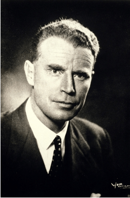
Professor Robert E Kendell was the senior consultant on Ward 2 and occupied the Chair of Psychiatry at Edinburgh University. He would go on to become Dean of the Faculty of Medicine at Edinburgh, President of the Royal College of Psychiatrists and eventually Chief Medical Officer for Scotland. He was an impressive man of conservative appearance and a somewhat unnerving presence. His inaugural address considered whether you needed a medical degree to treat mental illness. His conclusion was that if you were going to prescribe drugs or treat conditions that were the result of degenerative changes in the brain, you did. However if you were purely acting as a psychotherapist, you didn’t. He was pragmatic and used electroconvulsive therapy (ECT), where appropriate, without hesitation. I witnessed its transformative effects in very severe depression. All trainees were expected to administer it (to the non-dominant hemisphere) assisted by an anaesthetist.

My first patient interview was with a young male paranoid schizophrenic. He had been visiting brothels in town and as his illness progressed he had been involved in some violent behaviour which brought him to the attention of the police followed by his admission. As recommended in the textbooks I asked him how he was feeling. After a pause he said, ‘You look like a giant.’
‘And how does that make you feel?’ I asked.
‘It makes me feel like killing you.’
Alone in the room with him I glanced at my watch. ‘Well, I think it’s time for lunch,’ I offered cheerfully. ‘Why don’t you pop back to your room?’

Another early case was Lawrence, an elderly Jewish man who was admitted with depression. He had no appetite and had become emaciated. The skin in his axillae was deeply pigmented. I was quite convinced he had advanced malignancy and presented my diagnosis to Professor Kendall. He himself was a former physician, a neurologist, but made great show of deferring to my more recent qualifications. I thought he might be mocking me. Reluctantly he allowed me to transfer Lawrence to the Royal Infirmary for investigation, while he himself remained convinced the problem was psychotic depression. About two weeks later Lawrence returned from the Royal, the physicians having found nothing physically wrong with him. ‘Now will you let me treat his depression?’ asked Kendall with a twinkle in his eye. Lawrence eventually went home after a course of ECT completely recovered and back to his normal weight. Even his pigmentation resolved.

Unfortunately as the year wore on we trainees were given only minimal supervision and the lecture course seemed random and impractical. Nevertheless we were expected to see and manage both in- and out-patients ourselves. The nursing staff at least were helpful and pragmatic. An acute psychiatric ward is a very hands-on place to work. It takes enormous skill and patience to manage such challenging patients. On the morning housekeeping ward rounds with the nursing staff there was no point labouring technical diagnostic points. You needed to know how things were going – but briefly. ‘How’s John then?’ would be greeted by, ‘Still very mad.’ There was no need to say more. But this practical approach could be misinterpreted. A new social worker straight off a university course arrived on the ward. She was full of good intentions and theory but no experience. After her first ward round she turned to me and said, ‘I look around this place and I ask myself, who is giving these patients love?’

I loved the elderly secretary on Ward 2. Apart from being a first rate source of gossip, she took shorthand then typed up my letters, improving my prose style in the process. It was my only experience of dictating to a human being, all other jobs made use of small hand-held tape recorders. As she scribbled away, her cigarette never left her mouth and the smoked curled up through her glasses leaving her brow and forelock lightly tar-stained. She knew everything about the Royal Ed and the psychiatrists who worked there going back decades.

Arch humour was the norm among the experienced nursing and medical staff. It often took the form of a feigned callousness or deliberate contrariness. I claimed that if you said good morning to someone at the Royal Ed you’d be asked what you meant by that. One aphorism circulating in 1983 was that ‘neurotic’ had become a term of endearment. It was being applied to almost everyone and hence had become quite meaningless.

That Christmas of 1983 there was a Ward 2 night out to the notorious Armenian Restaurant in Holyrood where the highly eccentric owner insisted we all get up and dance to Armenian folk music. RE Kendall got very enthusiastic, attempting a sort of Cossack dance and ended up crashing into the stereo system.

Studying psychiatry was a social minefield. Asked what you did for a living at a dinner party the response to the information that you were training in psychiatry was unpredictable. Your dining companion might launch into a prolonged history of their sibling’s mental health issues assuming you’d be fascinated – or else they might become hostile because of some previous negative experience or philosophical dislike of the specialty. ‘Who decides if someone is mad? I mean, what would happen if you decided I was mad?’ was a common challenge. I would try to explain that the patients I dealt with were mad in the colloquial sense. A few brief moments in their company would convince any lay person of their insanity. They were obviously mad, not matter-of-opinion mad. I started saying simply that I worked in a hospital.

Within a few weeks I had become convinced that psychiatry was the wrong career move for me. This disaffection added to a background dissatisfaction with life and relationships in general. A deep gloom descended. I decided to look elsewhere for a job as soon as the opportunity arose but in the meantime I carried on in case I was mistaken and simply suffering from my own little unhappy psychiatric disorder.

Ward work and lectures continued. I began to get the hang of dealing with psychotic patients. Once they recovered they usually wanted nothing more to do with you, even crossing the street to avoid you as if merely being seen with you in public was humiliating. One patient in particular illustrated this problem.

Keith was a clever boy from a modest social background, the first of his family to go to university, and his parents’ pride and joy. After starting the first year of his physics course at Edinburgh University he experimented with marijuana at a party. This seemed to trigger his first bout of psychotic illness. It took the form of what used to be called schizoaffective disorder, a psychosis that combines features of schizophrenia and manic-depressive psychosis. Keith formed the conviction that he’d discovered the Fifth Force in the universe. He was unable to sleep.

Clearly unwell, his parents had taken him home from university but shortly after that he interrupted a physics class at his old high school, one that contained his younger brother, and addressed the class on the subject of the Fifth Force. The physics teacher didn’t like to stop him, perhaps imagining that like the sleepwalker myth it would be a mistake to interrupt him. Having come to the attention of the mental health services in this way he was admitted to the ward. His parents were very unhappy about it, feeling it would damage his career prospects. Eventually they demanded his release from the ward. Reluctantly we agreed, but his symptoms deteriorated further culminating in him vandalising the family car after which he was readmitted, this time under Section 28 of the Mental Health Act 1959.

Keith was ill for many weeks. His parents were devastated. When I arrived on the ward in the morning he would shout his delusional ideas in my ear and demand his release as I walked down the corridor. Each night I prayed, ‘Please make Keith better tomorrow.’ Eventually the storm subsided. Some insight crept into his conversations with me and finally he was released to out-patient follow up. He eventually defaulted from review, but on his last visit to out-patients he informed me that he didn’t believe any of the things he said to me while on the ward. ‘I just said that stuff to wind you up. I was laughing at you,’ he said, scornfully.

Meanwhile, my cohort of out-patients with neuroses began to grow. Every time I suggested they might be well enough to return to their GP for onward care they threatened to harm themselves. On one of the rare occasions I got to discuss this with my consultant he told me I was ‘encouraging dependency.’ My archaic medical model-type thinking tripping me up again.

III

However unhelpful, the lecture course was at least interesting. One highly anticipated session was the annual talk on hypnosis given by Professor Ian Oswald. The rumour was that he would attempt to hypnotise the whole class of trainees. Exciting stuff. Came the day and the Prof’s introduction:

‘Now, we all know that it’s easy to make fun of things and have a bit of a laugh, but we will never learn anything with that sort of approach, so I would like you all to take what is to follow seriously so that we may benefit from the experience.’ A frisson ran round the room. I was sitting next to my old university friend Jamie, also on the training scheme. Prof Oswald continued at a brisk pace. ‘Would you all now put both your arms above your heads and clasp the fingers of your hands together as hard as you can. That’s good. Now keep pressing as hard as you can. I’m going to suggest to you that when I tell you to unclasp your hands you will be unable to do so, no matter how hard you try… OK, now unclasp your hands.’

Jamie and I and most of rest of the class immediately released our fingers and looked around, relieved to have retained control of ourselves. However, a couple of our number were stuck with their hands locked together. Prof Oswald homed in on his victim, a quiet, diffident female colleague. He told her she would now be able to release her hands, which she did, then he told her to come to the front of the class. He sat her on a chair facing us and placed her in some kind of waking trance. He demonstrated a number of subjective phenomena such as telling her she could feel nothing on one side of her body. Finally, he woke her up and asked her if she thought she’d been asleep. She said no she hadn’t, she had been fully aware during the entire experiment but unable to do other than comply with his instructions.

Hypnosis as a public entertainment was legal in Scotland. Some of these performers offered other services such as cessation of smoking. One client became suspicious of the therapist’s motives when he suggested they meet at her home. Her brother-in-law was a policeman and arranged for officers to be present secretly inside the house. Although the hypnotist was arrested for attempted sexual assault and came to trial he got off on the basis that the police tactics amounted to entrapment. Prof Oswald was an expert witness at the trial. Fascinating stuff.

A less enjoyable incident occurred during a lecture on psychotherapy from Professor Henry Walton. Walton is now famous for his modern art collection, donated to Edinburgh galleries. At that time he was better known for his thoughts on the suitability of medical students to do psychiatry based on their ‘tolerance of ambiguity.’ He had authored a popular science book called Know Your Own Mind. I remember a lecture he gave to us as medical students which involved him taking a sexual history from some poor man in a dressing gown in front of a large audience. The man stared at the floor throughout as Walton asked about masturbation etc. and didn’t answer him. ‘Do you always experience such difficulty discussing sexual matters?’ asked the professor. I did a cartoon of this appalling scene and questioned whether I wanted to be a doctor at all.

Walton was notorious within the medical school for an episode of shoplifting in Harrods which he eventually attributed to a fugue state. When asked by the trial judge what a man in his position was thinking of being caught shoplifting he said, ‘I don’t know, I must have been mad.’ Private Eye pounced on this exchange. A period of gardening leave followed, incompletely observed, then he was reinstated. By the time I attended this lecture I was battle scarred and thoroughly disillusioned with the training scheme. At the end of his exposition Prof Walton invited questions. I raised my hand.

‘Professor, we are very junior trainees who have only a few months of experience but on-call we are invited to see acute patients and offer short term psychotherapy if appropriate. They often have massive thick notes indicating they have seen many, more experienced, psychiatrists in the past. I’m afraid I feel a bit of a fraud offering my services to them.’ The professor’s face darkened, ‘If you said that to me in an examination I would fail you and trust you would take up some other branch of medicine.’ I thanked him for his reply.

About a week later I bumped into Chris Freeman one of the other consultants on Ward 2. By this time I had moved to Ward 4 for my next six month attachment.
‘I wanted to speak to you Allan,’ he began.
‘You want to know how I’m getting on in Ward 4?’ I offered cheerfully.
‘No, I don’t care about that,’ he said (more psychiatric humour) ‘I wanted to talk to you about what the Prof said to you in the lecture last week.’ Somehow the exchange had come to his attention.
‘I want you to know that it will have no bearing on your future career in psychiatry.’
‘No I suspect it won’t, but thanks for telling me,’ I replied.

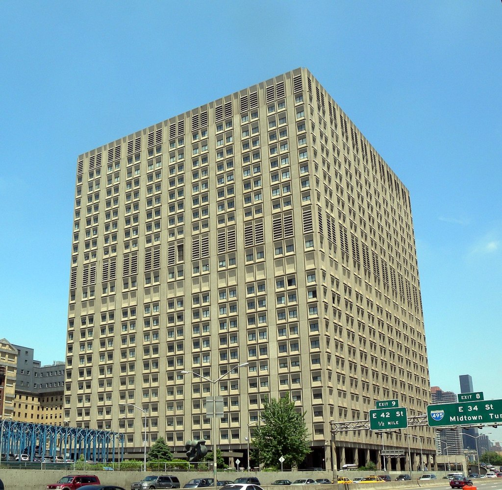
On-call work at the Royal Ed could be challenging. Two trainees were available on any given evening, one was based at the Royal Ed itself. After 10pm the other trainee would make their way to Craighouse Hospital, a spooky Victorian long-term asylum on a nearby hill that looked more like the set for a horror movie. It had originally been built to house wealthy patients and their servants in gothic luxury but had since become an NHS facility. Sleeping there and hearing the ‘noises off’ was unsettling but at least you weren’t at the mercy of acute admissions.

Back at the main hospital you waited to be summoned. There was a mixture of outside referrals and in-patient crises to deal with. One night before my colleague had left for the Addams Family Mansion I got a phone call from switchboard. I had a call waiting. A weary-voiced woman informed me that she thought she’d killed her baby. She refused to tell me who she was or where she was calling from. In the background a large dog barked incessantly. Despite the information that ‘the wean’s no’ breathing’ the woman wanted to discuss other issues. I covered the mouthpiece and whispered to my colleague, ‘Can you trace this call?’ She shrugged then picked up the other phone in the room. Eventually the woman became impatient with my amateurish advice and rang off. ‘You’ve been nae help tae me!’ she chided. My colleague told me the police had traced the call.

Half an hour later the police called me. ‘We’re at the property now doctor. The door’s locked and we can hear a dog barking inside. How should we proceed?’ I was well into uncharted waters now. ‘I don’t know. You’re the guys on the spot.’ I said. ‘All I know is that she claimed to have killed her baby.’
‘OK, doc we’ll let you know what happens.’ They rang off.

About 15 minutes after that they were back on the phone. ‘Eh, we broke down the door and there was just a dog inside the house, no baby, and now the owner’s come back from the pub. She’s very unhappy about the damage to her property.’

In-patients would often harm themselves overnight. At times basic first aid was required but often the damage was more serious. A rugby player, a front row forward with a local club, was admitted with his first psychotic illness. The ward called me to say he had stripped himself naked and was jumping off his bed onto the tiled floor head-first because, ‘God is telling me to have epileptic fits.’ The burly charge nurses from the high security Ward 10 were already on the case and I was required to complete the formalities.

When I got there the man seemed calm and I began completing a Section 28 and countersigning the drugs that had already been administered to the patient by the nurses. The patient and his escorting posse walked down the ward and out of sight towards the stairs. Suddenly a young female nurse came running back towards the duty room shouting for me to come quickly as there had been ‘an incident.’

The patient had made a break for freedom at the top of the stairs, the nurses had restrained him and hauled him to the floor. In bare feet, one foot had stuck on the tiled floor as they piled on and he had sustained the worst fracture dislocation of his ankle I had ever seen. The foot was up one side of his tibia, the skin stretched tightly over the bone end. Despite this appalling injury the prop forward was still trying to get up! More sedation was given and I hurried off to phone casualty at the Royal Infirmary. ‘I’ve got an interesting one for you,’ was my opening remark.

By 4 am after several other calls I had fallen asleep in the horrible on-call room. There was a plastic sheet under the actual sheet which made it slide about in an unpleasant way and the big cast iron Victorian radiator was broken, stuck at maximum flow. The room was sweltering and you had to open the window wide to try to cool off. The phone rang. It was Ward 10. ‘Can you come and write up some analgesia for your patient with the broken ankle?’
‘What? He’s not at the Infirmary?’
‘They bivalved his plaster and sent him back. He’s OK, it’s just that he’s awake now and in quite a lot of pain. He’d had so much sedation before he went over there they didn’t give him anything else.’

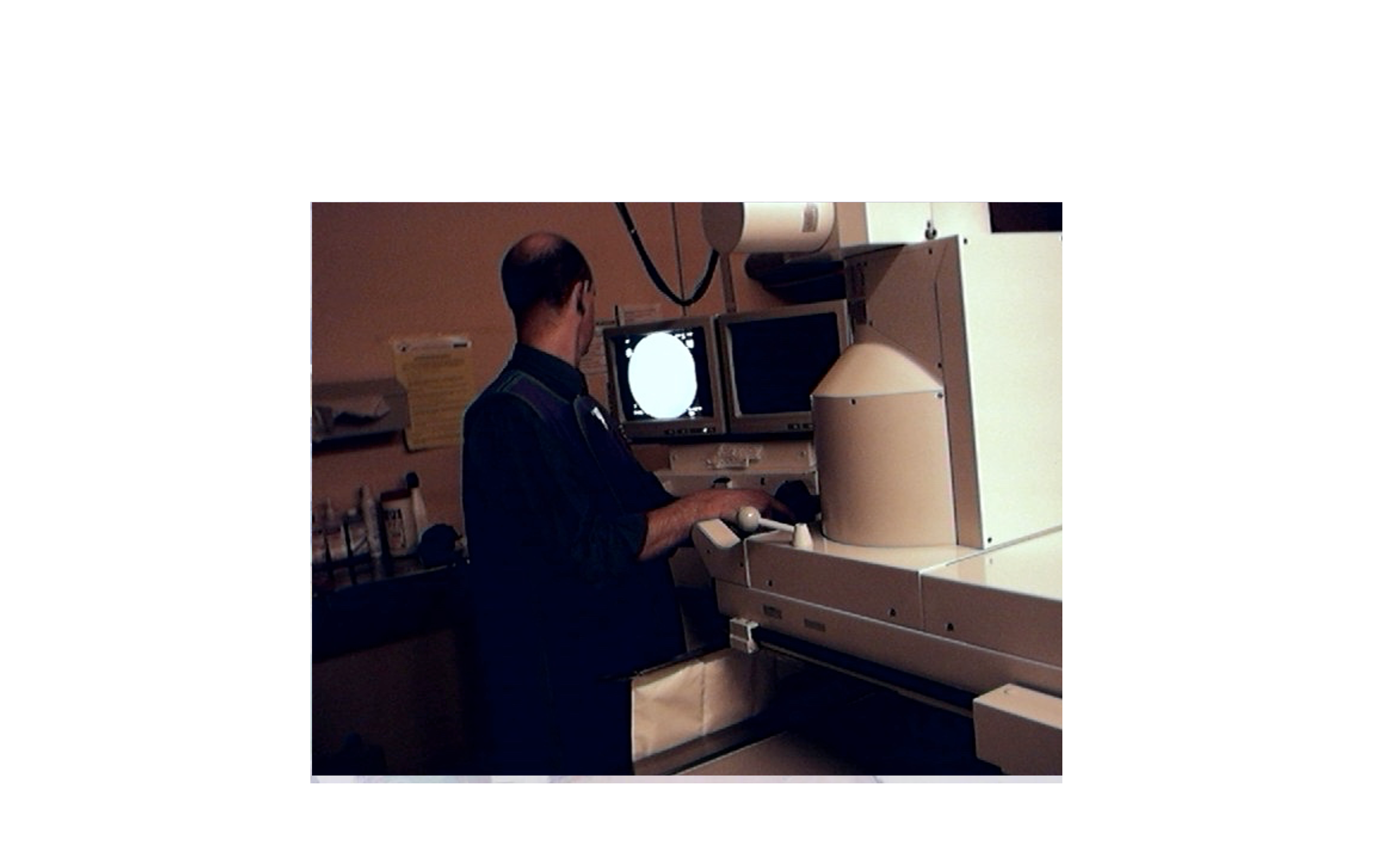
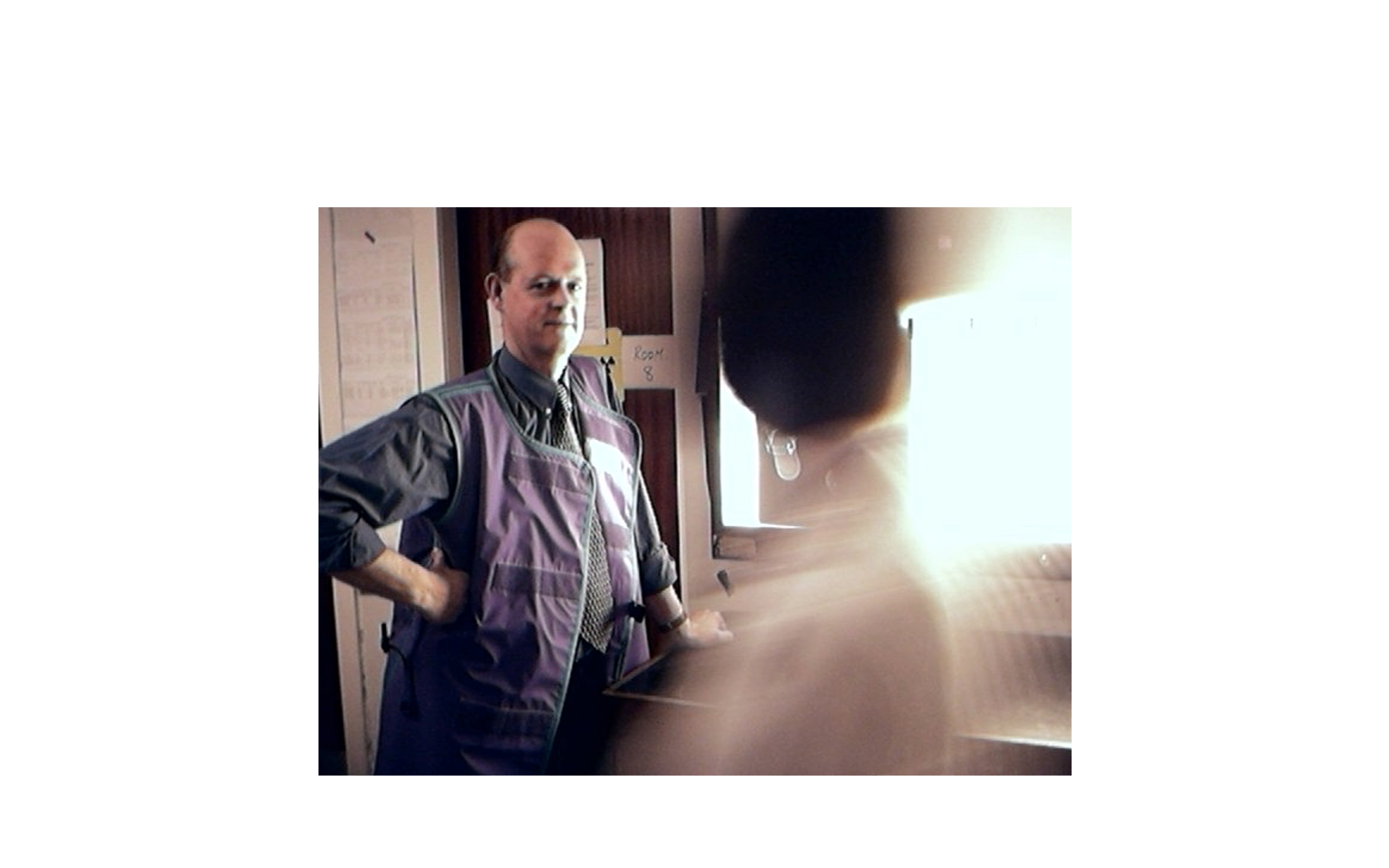
After Christmas I was still miserable and wanted to escape from this first career misstep. Seeking certainties I applied to the radiology scheme and was accepted. I decided to complete the full year in psychiatry rather than bail out to some other temporary job. After all, I might change my mind.

I completed my year in psychiatry with six months in Ward 4. The senior consultant there, whom I won’t name, was less helpful than REK. Once when reminiscing about his career he said, ‘At the time I had a number of sexual skittles in the air,’ a phrase that I’ve never forgotten. It proved pointless asking him for help with my now huge out-patient load.

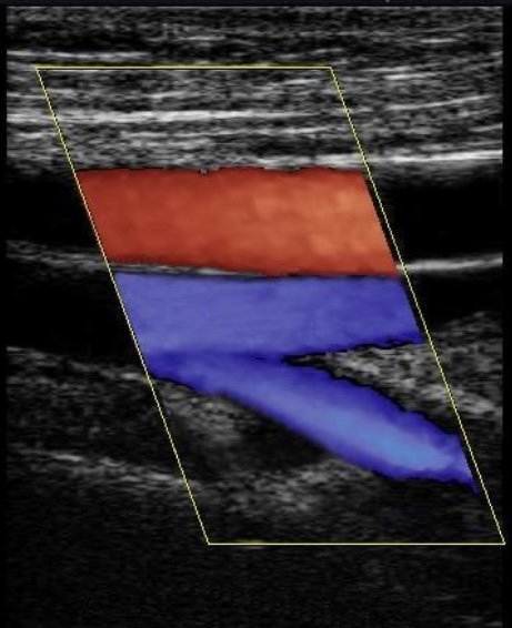
Towards the end of the Five Nations rugby tournament of 1984 Scotland found themselves in a Grand Slam decider with France at home. My brother got us tickets at the last minute but I was on call that weekend. In the registrar’s room I spotted the new trainee who had just arrived from Singapore. I asked him if he would swap on-call, and having nothing to do and no interest in Rugby Union, he agreed. I got to stand on the North Terracing and witness a Scottish victory. Jerome Gallion the French scrum half was knocked unconscious in a collision with David Leslie and admitted to the Infirmary for a CT scan in the department I was soon to join. It was the only time I’ve seen supporters throw their hats in the air.

Exeunt

When Jamie and I announced our departure from the scheme there was a certain amount of reflection on the part of the trainers. We were asked to explain ourselves at the Friday afternoon ‘sensitivity meeting.’ Having secured new jobs we did not really want to explain ourselves and instead blamed personal issues such as our emotional unsuitability to the cut and thrust of mental health. Beyond that Friday afternoon session I was summoned to Professor Kendell’s office to explain myself. He seemed disappointed in me and I felt uncomfortable. He plonked me in a very low armchair then sat perched on his desk high above me.

‘Why are you leaving us, Allan?’
I struggled to form a sensible response. ‘Well, I find the patients awful,’ I offered lamely, reluctant to say that I felt completely unsupported.
‘What’s awful about them?’
‘Well they seem to get better or worse irrespective of what I do for them.’
‘You don’t find them interesting in themselves? You will be aware that before psychiatry even existed as a science the great descriptions of psychological illness were found in literature; in Shakespeare and Dickens. What interests you in medicine?’
‘Structure and function I suppose,’ said I.
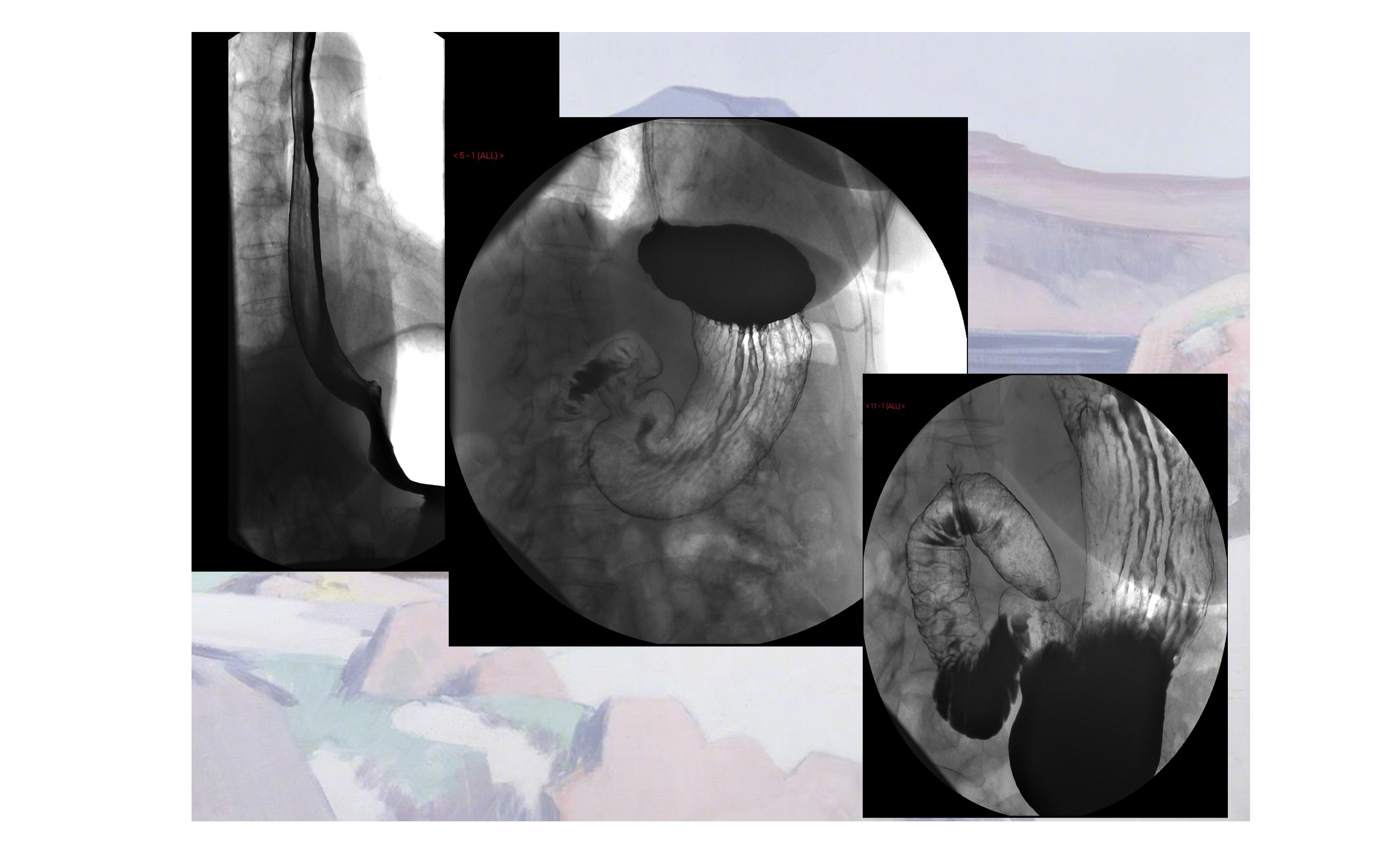
‘Well, we could be a hundred years away from that in psychiatry,’ said Kendall. ‘And why radiology for goodness sake? Radiologists lead an etiolated existence.’ He sighed. ‘Write to me and let me know how you get on.’ With that I was dismissed and went home to look up etiolated.

After a few months of happy structure and function in radiology – the plumbing and wiring of medicine – I decided I should indeed write to REK and tell him that I was satisfied I’d done the right thing. I sent him an anodyne note thanking him for his counsel and the reference he had written for me.

A few days later I got a reply:

Dear Allan,

I do hope you settle down in your chosen metier soon. Should you have any doubts about your decision I suggest you recall what you inscribed on one of our lecture theatre desks not six months ago: Psychiatry is bunk. AJMS 83-84.

I trust this was an accurate reflection of your feelings at the time.

Robert E Kendall.

I was horrified. When I told his friend Judy Greenwood about it she dismissed it, ‘Oh he’s a bugger,’ she laughed. ‘I’ll have a word with him.’

Years later when I had become a consultant radiologist I discovered REK had been taken off my ultrasound list by a senior colleague who felt I wouldn’t want to scan my ‘old boss.’ Some time after that I met him leaving outpatients and he recognised me. ‘You had a lucky escape Prof. You were nearly on my ultrasound list.’ I said, grinning. ‘You’re the one who missed out,’ he replied. ‘I am an interesting case.’

Becoming a Haruspex II: Ultrasound

A third generation Scottish doctor, Ian Donald was mentioned in dispatches during the war after he pulled several airmen from a crashed and burning bomber. The bombs were still on board. As Regius Professor of Obstetrics and Gynaecology at Glasgow University in the 1950s, he thought industrial ultrasound scanners might be adapted for use in human subjects. They were. There is a famous picture of him scanning his pregnant daughter. At the end of his illustrious life, he worked for a while at the Western General in Edinburgh. I was told he estimated how much diuretic he needed to take for his cardiac condition by scanning his inferior vena cava in the morning.

https://en.wikipedia.org/wiki/Ian_Donald

Technique

A back-of-an-envelope calculation indicates I have performed ultrasound scans on about 40,000 people in a 33 year-long radiology career. This would be a decent gate for a sporting event and enough material to have seen most things ultrasound can reveal. I did at least two ultrasound lists per week of up to 25 patients plus ad hoc scanning at other times. A list had to be completed in under four hours, which left no time for slacking. Ideally, you set up two ultrasound rooms with an ultrasound ‘helper’ to get the patients onto the couch and ready to go. You could then move rapidly between the rooms and maximise your scanning time. Having a junior trainee with you whose scans needed to be checked slowed you down considerably. Underlying all this intense activity was the necessity of getting it right.

You need experience to interpret what you are looking at on an ultrasound image. To the tyro, the ability of a senior radiologist to recognise pathology instantly seems almost psychic. With common conditions the diagnostic experience is akin to recognising an old friend; with rarer ones it’s more like meeting a celebrity. The secret to success is putting in the hours – and maybe a little bit of aptitude. You can learn to play a piano if you can learn to type, but genuine musicality needs talent.

In the second year of our radiology training we were allowed to start doing ultrasound. To me, the process seemed magical and it remained my favourite technique. The silly description of it as ‘putting jelly on the belly and watching the telly’ degrades the wonder of it all. Like a magic wand, you pass the probe over the body and a fan-shaped image of the organs lying under the skin appears in real time on the monitor.

An ultrasound signal will not cross an air gap. The probe needs a coupling medium to both conduct sound into the body and to receive the returning echoes. Early techniques used a water bath, but ultrasound gel is a lot more practical. The time it takes for the echo to bounce off a structure and return to the probe places that signal at a specific depth and from that data images are constructed. The physics and electronic engineering of all this is very complex. Modern probes have no moving parts; multiple piezoelectric crystals are arranged in an array in the head of the probe. These crystals emit ultrasound and respond to the returning echo by emitting a tiny electric pulse.

The triangular image seen on the monitor tapers upwards towards the position of the probe on the skin surface and is rendered in monochrome. You can scan the patient at any angle you please. Scanning in the sagittal plane – the long axis of the body, front-to-back – shows you the organs from the side. Similar to the air-filled spaces outside the body any air present in bowel will also interrupt the signal and prevent visualisation of any structures behind it. So will bone, which is impenetrable to ultrasound. Like bone, the stones found in fluid-filled organs like the gallbladder and urinary bladder will reflect the sound beam and cast a diagnostic dense ‘acoustic shadow’ behind.

A gallstone

The higher the frequency of the sound, the greater the detail displayed but higher frequencies penetrate less depth of tissue. For that reason very high frequencies can only be used to scan superficial structures such as the eyes, the thyroid and the testes. The latter organs might well have been designed for ultrasound lying – as they do – outside the abdomen. The image resolution within these small superficial structures is remarkable, often described as ‘exquisite’ for some reason. Demonstrating larger structures like the intra-abdominal organs demands a lower frequency and some detail is inevitably lost.

When you first sweep a probe over a patient’s body there is a tendency to look at the probe or at the patient rather than the monitor display. You must learn to do the physical part of the scan automatically while concentrating on the images on the screen, similar to looking through the windscreen while driving a car.

I usually did a quick survey of the abdomen to get the lie of the land before scanning the patient in detail. Once, while doing a pelvic scan on a young woman, I noted a mature follicle in her right ovary. This indicates that ovulation is imminent,. That happens in an instant as the follicle ruptures. I scanned her uterus in the midline then across to her left ovary before returning to the right side where I noticed the follicle had started to collapse.
‘Do you ever get pain at mid-cycle?’ I asked her.
‘Yes,’ she said. ‘In fact, I have a bit of a pain at the moment…’
Mittelschmertz (middle pain) is the medical term for the discomfort of ovulation.

Since the images generated are very thin two-dimensional slices you must learn to construct a mental picture of the organs you are scanning based on your anatomical knowledge. The three-dimensional structure of the patient’s liver, biliary tree, gallbladder, spleen, pancreas, kidneys etc is built up in your mind as you scan through them – like shining a torch around a darkened room and remembering what you’ve seen. Actual 3-D reconstructions of ultrasound data produce a confusing jumble of echoes too difficult to interpret and if you surface-render ultrasound it takes you back to square one. After all, the patients themselves are ‘surface-rendered’ structures that you cannot see through. The popular 3-D images of fetuses impress the lay person but are of limited diagnostic value. The concept that thin slices through a structure are easier to interpret than a 3-D image is difficult for non-radiologists to grasp.

Turf Politics

Clinicians would frequently ask me what the ‘trick’ was to ultrasound. I always replied that if you turned the film upside down the answer was written along the bottom. Obviously, the actual ‘trick’ is to do thousands of scans. Some images, particularly any measurements, are recorded for clinical record-keeping and as an aide memoire when reporting, but the crucial thing is your opinion rather than those tricky scan images. Clinicians have to trust the accuracy of your report, which is where the issue of confidence in an individual radiologist arises. Clinicians like to know which radiologist has reported their patient’s scan and they like to have critical scans discussed in joint meetings for clarity. Referral to a specific radiologist is very common in private practice.

After about two years of doing hefty ultrasound lists you begin to get the hang of it, and good enough to fly solo. For non-radiology-trained clinicians to become similarly competent would require them to undergo the same amount of training. Despite this, clinicians would sometimes have a crack at doing ultrasound themselves. They would get hold of some ‘soft money,’ buy their own scanner, then go on a weekend course at a hotel somewhere funded by the ultrasound manufacturer. As Arthur Conan Doyle once said, ‘mediocrity recognises nothing above itself’. A lack of insight into your limitations can result in mis-diagnosis and potentially harmful consequences for the patient.

Many years ago the general surgeons in the main hospital where I trained decided that they didn’t want to bother with radiologists any more. They felt they should crack on with doing their own scans. The consultant in charge of our service said that if they did that he wouldn’t be doing any more scans for them. A threat he would be unlikely to carry out. After the usual weekend course they bought their own scanner. They did not ask for any training from us. On the basis of ensuring safe medical practice we would have refused anyway. We had enough on our plate training our own juniors. 

Some time later, on a Monday morning, the surgeons came down to the department and asked to speak to the boss. They were puzzled. They had detected gallstones in a patient at the weekend and taken them to theatre only to find that the gallbladder was quite normal. My consultant reluctantly agreed to look at their images.
‘See,’ they said, ‘There are the stones.’ 
‘Well, actually that’s the right kidney,’ replied my boss. 
The surgeons stopped doing scans after that and abandoned their scanner. It is very wasteful to have a nice new US machine sitting in a corner under a dust sheet doing nothing. That does not happen in radiology departments.

An impatient but charming gastroenterologist once stuck his head round the door of the scanning room to ask if I’d do an urgent abdominal scan for him. At the time I was examining a baby’s head through the fontanelle (‘soft spot’) looking for any intracranial haemorrhage. When I spoke to him later he asked me what I was doing to the baby and was surprised to learn that US had this application. Out of devilment I asked him why he didn’t do his own scans to which he enquired how long it had taken me to become proficient at it. I said it had taken two or three years of scanning several lists a week. ‘Well, I don’t have the time to do that!’ he said. ‘I don’t do my own biochemistry and I don’t intend to do any ultrasounds. I prefer to get you to do it and send the patient back to the clinic with the right answer.’ As a gastroenterologist skilled in endoscopy, he had plenty of procedures of his own to do. Ironically gastrointestinal endoscopy has almost completely replaced the barium studies that were once fundamental to radiology practice – and a huge part of my own early training. The relentless advance of technology has swept many skills into the dustbin of medical history.

This is precisely what happened when lower limb venography was replaced by ultrasound. A swollen or painful lower limb can be the result of a deep vein thrombosis (DVT). Most people are familiar with this as a complication of long-haul flights, but DVTs also occur in a variety of other clinical settings. The danger is that a clot that has formed in a leg or pelvic vein suddenly becomes dislodged and travels north, through the right heart and into the lungs. If the clot blocks the pulmonary arteries oxygenation of the blood fails and the consequences can be fatal. Smaller clots cause breathlessness and lateralised chest pain aggravated by breathing. Venography had been the way to detect blood clot for many years. It was the ‘gold standard’ of DVT diagnosis and clinicians were confident in it.

Lower limb venography is the injection of X-ray contrast into a small vein in the foot so that the larger deep veins of the legs and hopefully the pelvis can be demonstrated on X-rays. It produces hard copy in the form of radiographs that show recognisable veins. Ultrasound images on the other hand make no sense at all to clinicians, requiring them to depend entirely on the opinion of the radiologist. This makes them uneasy. The appeal to us was getting rid of venography – a difficult and unpleasant examination – and replacing it with ultrasound. Of course it was necessary to demonstrate that in addition to being safe and painless, ultrasound was at least as accurate a technique as venography.

Venograms were by definition always urgent and seemed to gravitate towards Friday afternoons. Swollen feet can make finding a vein very difficult. Touniquets were applied above the knees and ankles. If necessary the patient’s feet were heated with warm towels or a basin of hot water to encourage venous engorgement. Any swelling present had to be pressed away using your thumbs. Once you had established venous access the patient was positioned on an X-ray table ready to take films as the contrast was being injected. About 100 ml of contrast was injected into each foot as fast as you could manage. I used to hold the syringe barrels in both fists and force the plungers against the front of my lead coat. You hoped the rapid flow of viscous contrast would not blow the veins you had punctured. Two views of the calves were followed by blind films of the thighs and pelvis as you removed the tourniquets and lifted the legs up to encourage flow. The contrast would then travel up into the abdomen. There was some skill in knowing how long to wait before telling the radiographer to take the film.

We were keen to get rid of this awful test – but the clinicians were resistant. We presented our case at hospital ‘Grand Rounds’ supported by our own research and papers from the the world medical literature. Our research had involved doing a venogram and an ultrasound on every patient referred for venography to show that we were not missing any significant thromboses. We emphasised that we could also pick up other conditions on ultrasound that masquerade as thrombosis – like ruptured knee joint cysts. Venography could not detect these. Eventually we convinced our colleagues and succeeded in replacing the venography service with ultrasound. Ironically, clinicians soon perceived that US was a reliable benign, non-invasive procedure compared to venography and their threshold for requesting tests dropped!

Some specialties, cardiology for example, are limited to just one anatomical area. It had become clear to cardiologists that they would need to learn ultrasound of the heart and angiography of the coronary arteries to avoid simply becoming a referral service for radiology. All of that was inevitable, but without general training non-radiologists would not recognise other conditions lying outwith their own particular area of expertise.

The Passage of Time

The biggest change in ultrasound practice in my working lifetime was the emergence of radiographers, the radiology technicians, as diagnostic ultrasonographers. That role had previously been the preserve of the medically qualified radiologists. The pressure on radiologists to deal with the massive increase in workload from the new cross-sectional imaging techniques (CT, MRI and PET) had encouraged this trend. The British Medical Ultrasound Society is now largely a radiographer-led organisation.

Unlike the reporting of disembodied X-ray images, when you do an ultrasound you have a real live patient in front of you. You can take a history from them and even exchange pleasantries. After a glance at the clinical information on the request form (not always helpful) you scan what lies beneath. As things are revealed you can ask the patient questions in real time to help clarify the diagnosis.

Sometimes a returning patient would ask me what the scan was showing.They had grasped that you were making that decision at the time, as you would when feeling their abdomen or listening to their chest. ‘I hope you’ve got good news for me today doctor,’ was a common greeting as the patient entered the scanning room. I would never mislead anyone, but as I was not in charge of their ongoing care, imparting bad news to a patient could be problematic. At least if they were going directly to their clinician after the scan any issue could be promptly dealt with. An oncologist once told me it was ‘helpful’ if I’d given the patient the impression that all was not well. I felt that was actually their job.

Despite all this I looked forward to ultrasound sessions, even though they were invariably over-booked forcing you to work at an uncomfortable pace. I used to compare it to driving down the high street on a busy Saturday morning at ever increasing speed. How long before you miscalculated and hit a pedestrian?

Beyond the purely diagnostic work lie interventional ultrasound procedures. This is the use of ultrasound guidance to carry out treatments that would have previously required some form of conventional surgery. Examples would be the targeted biopsy of small, deep-seated tumours or the drainage of an organ or fluid collection. Surgeons have the advantage of an anaesthetist to put the patient to sleep and look after them while they do their thing. Doing this type of work as a radiologist meant managing the understandable anxiety of a conscious patient while simultaneously performing a tricky procedure under local anaesthetic. You could give intravenous sedation – but if the patient nods off, you’ve given them a general anaesthetic. Adequate analgesia is crucial if you are going to penetrate sensitive tissues with a needle or a drainage catheter.

Anecdotage

I once had to drain both kidneys in a very unwell patient and insisted on anaesthetic assistance. The disincentive is the delay that introduces to proceedings and the fact that the on-call anaesthetist may be busy elsewhere. By coincidence the anaesthetist who turned up was the same one who had done my wife’s epidural during the delivery of our second child. At the time I didn’t volunteer that I was a doctor, not wishing to put him off his game. He’d been rather tetchy, proclaiming that my wife would deliver soon enough anyway without the need for an epidural. At the time we were both quite annoyed with him. Once the kidneys were successfully diverted he complimented me on a promptly completed task and, frowning slightly, asked if we had met before. I explained that we had and outlined the circumstances. He asked if it had been a good epidural. I said it was and he smiled. After that we exchanged pleasantries when we passed each other in the hospital corridor.

The technical challenge of targeting small solid tumours in the deeper part of organs like the liver is considerable. The needle shaft is of uncertain visibility on ultrasound and, like a long shot, small errors at the outset can result in a miss. Core biopsy devices give a better specimen for the pathologist to look at but also put a bigger hole in the patient. I often took my own precious biopsy specimens along to the pathology lab to make sure I had hit the target – and to make sure they didn’t get lost.

A technique largely now taken over by radiographers is obstetric scanning. I learned to do these scans when I first took up my consultant post. The bulk of them are done to estimate the date of delivery but patients who experience early pregnancy bleeding need scanned for ‘viability’. A non-viable gestation means a difficult conversation followed by a trip to theatre. We were often contacted by GPs who wanted ‘a scan for reassurance’. I soon learned to ask the GP to tell the patient that if they’d had any bleeding there was a possibility that reassurance would not be forthcoming. In the end we insisted that these referrals went to the gynaecologists first for an informed discussion of the possible consequences before the patient attended for their scan.

Beyond simple dating scans routine scanning at 20 weeks was introduced to exclude fetal anomalies. As the radiographers’ training progressed, radiologists were no longer required to do the simpler dating scans. We were still called in to look at any potentially abnormal pregnancies the radiographers had detected. Obviously medicine is all about disease but doing simple dating scans in healthy mothers was a pleasure. When that evolved into only checking abnormal scans a lot of the pleasure went out of it.

Tyson

I was carrying out a routine anomaly scan in a small hospital I worked in as part of my first consultant post. The woman’s partner was in the room and was watching me closely. As I began scanning I became aware of the sound of a large dog barking menacingly somewhere nearby. I glanced towards the window.
‘I tell’t ye no tae bring Tyson,’ said the woman to her other half, annoyed. I carried on with the scan – which was normal. Although it is possible to detect the sex of a fetus it is not usual to offer that information. Apart from the possibility of making a mistake, there are some settings in which that information might be misused. If you were asked, it was easier to be sure the baby was a boy than a girl.
‘Can you tell tell whit it is doctor?’ asked the man. As it happened, I already knew it was a boy.
‘Well, yes. Are both sure you want to know?’ I offered. The man and the woman exchanged glances, Tyson continued to bark furiously in the background somewhere.
‘Aye OK then,’ said the woman.
‘Well,’ I said, smiling, ‘It’s a wee boy.’
The man erupted. ‘Aw, no anither laddie!’ he shouted.
‘Ah tell’t ye no tae ask!’ yelled the woman. ‘Ah tell’t ye!’

Technicolor

Colour Doppler

The exception to US yielding monochrome images is colour doppler. The doppler effect is that drop in pitch you notice when a noisy car passes you at speed. Rapid approach compresses the sound waves raising the pitch then, as the car departs, the sound waves are pulled out and the pitch drops. The same effect is produced by bouncing sound waves off moving blood. Simple doppler samples a limited volume within a blood vessel but multiple measurements can be made across a whole image and converted into colour signal superimposed onto the black and white image. By convention red is ascribed to movement of blood towards the probe and blue away, but this is arbitrary. The intensity of the colour can be made to reflect the velocity of the blood. Turbulent flow jumbles the colours and narrowing of arteries produces a jet of very high velocity.

I gave a workshop in Athens to the Greek Ultrasound Society Annual Meeting. A lot of physics was involved and I tried to spice it up a bit. As I waited to start my talk, which was to be illustrated by videos of colour doppler examinations, I ran a clip of Celtic and Rangers scoring against each other in quick succession during a recent Old Firm match. This got their attention but blue versus green wasn’t a particularly helpful analogy. My later illustration of Scotland scoring a try against Wales at Cardiff at least involved the correct colours. ‘Red is movement towards us, blue away,’ I declared. The Greeks were unimpressed with Rugby Union. A few of them were asleep.

After an hour and a bit of me droning on I took questions. There were just two and both were about whether colour doppler might be useful in the investigation of erectile dysfunction. At the time I didn’t really know. My colleague later said this reflected the pattern of private practice in Athens – no pun intended. I later found out that yes, colour doppler was useful in this condition and I even performed some of these tests myself. This involved injecting the offending organ with a prostaglandin-related drug. A certain amount of commitment was required of the patient in order to achieve a diagnosis. Priapism followed by extensive thrombosis was a potential side effect.

That evening, after my colourful lecture, a highly elaborate ceremony was held at Athens University. Choirs sang, music was played and they presented all the guest lecturers with a silver icon. Afterwards there was a grand reception with drinks and nibbles. It was December and quite chilly for Athens. There was a familiar odour about the place which for some reason reminded me strongly of my grandmother. I couldn’t place it until I realised that many of the women were wearing fur coats and the smell was of moth balls.

Iconic

Röntgen Redux

One morning while attending the 8:30 clinical meeting with the urologists I was called out to take a phone call at reception. It was from a physicist who was an expert in subsurface radar. I’d never heard of such a thing. He had previously worked for the military and had a laboratory out in East Lothian somewhere. He wanted to explore potential medical applications for his technique. So far he had used it successfully to find sunken ships in the Mediterranean and unexploded WWII bombs for the London Underground. He asked if he could come and see me to discuss a clinical trial. When I returned to the meeting a colleague asked who had called. ‘I think I might have been talking to the next Conrad Röntgen,’ I joked.

A couple of weeks later he duly appeared and I showed him my best ultrasound pictures. He seemed very disappointed in them. He could see ‘down to molecular level’ with his radar and was baffled as to how we could determine anything useful with such primitive equipment. I asked him if it would help to see some live imaging and he said yes, he thought it might. It was lunchtime and the list in the room with the best scanner had just finished. When ultrasound reps are trying to sell you their machines they often scan their own bodies as a demonstration, usually their abdomen or neck. I didn’t really want to get ultrasound gel on my abdomen or my clean shirt so I decided I’d use my neck. I would be able to show him colour doppler on my neck vessels.

Picking up the high frequency probe I put gel on it and stuck it on my right carotid artery. The first thing I saw was an atheromatous plaque near the carotid bifurcation. ‘Oh,’ I said, dismayed.
‘What’s the matter?’ asked the boffin.
‘I’ve got atheroma in my neck.’
‘What’s that?’ he asked.
‘It’s the thing that causes strokes and heart attacks,’ I said bleakly.
‘Did you know you had that?’
‘No.’

Needless to say the radar thing never got off the ground and we carried on with our ‘primitive’ techniques. The plaque in my carotid bothered me. I had looked at the other side, which was normal. Helpful colleagues told me that they too had looked at their carotids and were delighted to report that their arteries were all perfectly normal. I went to see a friend, a radiology professor in the neurosciences unit, because carotid atheroma was one of her specialist research subjects. She said carotid plaque was a common finding in 47 year old men like me but if I wanted to get my cholesterol checked I could. After a while I got my GP to do it and it was up. We checked it again a year later and it had gone up again.

Eventually I seemed to reach that part of the nomogram that indicated a statin would do me more good than harm and so I started treatment. I would occasionally sneak a look at my plaque to see how it was getting on. To my surprise it changed completely after I started simvastatin, becoming much more echogenic and possibly flatter. Of course, this may simply have been coincidence and part of the natural evolution of my arterial problem but studies suggest that atorvastatin, which I take now, can reduce plaque build up and stabilise it. Maybe I wouldn’t be sitting here typing this drivel if it wasn’t for a certain eccentric scientist.

Testing, Testing

I was referred a patient with breast cancer. She was the chatelaine of a grand Highland estate and had initially been treated in London. Her oncologist had asked me to scan her liver for metastases. We had no previous imaging in Edinburgh and no previous reports for reference. She swept into the room and addressed me with great confidence. A scan of the liver for metastases was a basic request. We performed such examinations several times a day. As I scanned her liver I picked up two small, intensely echogenic (bright), lesions in the right lobe. They were very well-defined and typical of benign haemangiomata, tangles of fine blood vessels. These are found in about 10% of middle aged subjects, particularly women, and are frequently multiple. They are the commonest type of benign liver tumour. I would not normally mention them to a patient but noting their presence in the formal report is important in establishing a baseline for later imaging. There were no lesions suggestive of metastases.
‘Well, are you finding anything?’ the grande dame enquired.
‘No. Well at least nothing you need worry about.’
‘What do you mean by that?’ she asked.
It was clear I would have to mention the haeamnagiomata. She would be going straight back to the clinic with my written report.
‘You have a couple of benign things in your liver that are of no significance. Nothing else.’
‘Would you say they were haemangiomas?’ I was surprised to hear her use the technical term.
‘Yes, that’s what they are.’
‘Good, that’s what they told me in Harley Street.’
She had been testing me.

Judging by Appearances

One of the benefits of doing private practice is the much more manageable pace of the lists. You actually have time to think and time for some bedside manner. Captains of industry can be tricky to deal with as they are not accustomed to taking anything on trust and have a tendency to interview you and make a judgement about your ability.

A few years ago a young man turned up for a testicular scan at the private hospital. He came into the examination room rather diffidently, dressed in a very smart pinstripe suit. He was tanned and I assumed he was a ‘young urban professional’ who had been somewhere expensive and sunny for a holiday. I greeted him and he nodded without saying anything. It is very common for patients attending for this examination to be highly embarrassed. I explained that he needed to loosen his trousers, get onto the couch then slip his pants down to mid-thigh level. Again, he said nothing. The scan was quickly performed and entirely normal. I handed him some paper tissues to clean off the gel.
‘That’s all fine,’ I said. ‘Everything is normal and I will send a report to your doctor.’ Again, he nodded but said nothing. He finished adjusting his apparel and I turned away from him to face the scanner and close his case, ready for the next patient.
‘So ye’re a ba’ man then?’ a broad Scots voice enquired.
‘Pardon?’
‘You’re a ba’ man. That’s whit ye dae?’
‘Well, it’s not all I do…’
‘Aye, we yase the ultrasound on the yowes, ye ken.’
I glanced down at the request card to see the address of a Borders farm.

Finally

I miss the magic of ultrasound. I have been scanned by colleagues and had the weird experience of lying on the couch wondering what they are seeing on the screen. In addition to scanning themselves, many younger colleagues scanned their wives during pregnancy, a practice that should properly be discouraged. I intended to avoid doing that with our children but during her first pregnancy my wife was concerned that she hadn’t experienced ‘the quickening’. In fact she had, but misinterpreted it as something else. This was all terra incognita to me.

Since she had already had a normal detailed scan I thought there would be no harm in having a recreational look at the baby. We went to the department one evening and as soon as I began scanning I saw there was an obvious cystic abnormality in one of the baby’s kidneys. The other kidney was normal as were the rest of the organs. There was urine in the bladder and a normal amount of liquor. The pregnancy was only 20 weeks duration at that point. We went home and I called a senior colleague. The next morning he confirmed the findings which had been overlooked at the detailed scan. 20 weeks of worry and further scans ensued before our healthy son was born – albeit with only one kidney. The knowledge of having a single kidney may be crucial to him in the years to come so I tell myself it wasn’t really the wrong thing to do. However, as one of my old consultants told me during my house jobs, ‘Never have a test unless you know what the result is going to be.’ Wise words.

Becoming a Haruspex I

In 1977 I went to New York to do a six-week student elective in gastroenterology at New York University. I arrived in the middle of a heatwave. The blackout that year had resulted in widespread looting; the Bronx was ‘burning’ due to insurance fires; the police were on strike and picketing the Brooklyn Bridge and David ‘Son of Sam’ Berkowitz, the .44 caliber killer, was shooting courting couples at random. His arrest for an unpaid parking ticket was imminent.

The placement was at Bellevue Hospital, a name forever associated with the notorious psychiatric wards. In fact Bellevue was also a general hospital housed then in an enormous brutalist block known as ‘The Cube’. The original infamous Bellevue building next door was being demolished while I was there but I managed to get into it and wander the deserted corridors looking for ghosts. When I heard some raucous voices coming from one of the abandoned rooms my courage deserted me. I fled back to the relative safety of the Cube with its handcuffed prisoners and armed police guards.

Bellevue was a public hospital paired with the private University Hospital known as UH. It lies at the east end of 28th Street between 1st Avenue and the FDR Drive. The UN Building is a few blocks to the north. Ward rounds started very early in Bellevue so that the attending physicians (consultants) could make their way along the underground passage from to UH – where the money was. In the UH lobby hung an Andrew Wyeth and a very expensive car showroom was close by. Despite these signs of opulence New York was in severe decline then.

The Cube on FDR Drive

Through an acquaintance with connections in Philadelphia I also spent some time in that city. It was noticeably less crazy than NYC. As a result I met someone who would become a lifelong friend. Al Dorof was living in an apartment on Delancey Place, a gorgeous mid-nineteenth century tree-lined street that has featured as a location in several films including Trading Places. It is amongst the most prestigious addresses in Philadelphia. The fabulous Rosenbach Museum of literary memorabilia is there. Delancey Place has hitching posts and mounting blocks and is older than many of the streets of Edinburgh’s New Town.

The palatial townhouse was divided into flats with a communal kitchen in the basement. Those Philadelphia contacts who introduced me to Al had prevailed on him to put me up for the weekend. We got on very well and I returned to Philadelphia to stay with him when my elective in New York finished. Also staying at Delancey Place was Larry, an acquaintance of Al, who was a radiologist.

One evening Larry came home from the hospital and said, ‘Look what I had to report this morning.’ He propped a frontal skull view against a lampshade and invited us to inspect it. There was a dense round structure projected over the exact centre of the frontal bone (the forehead). Then he showed us the lateral. A faint linear shadow could be seen traversing the skull from front to back, terminating in a triangular metallic structure just above the pituitary fossa. It was an arrowhead mounted on a wooden shaft. Along the line of the shaft tiny fragments of bone and pockets of gas were visible that had been carried in from the entry point. An endotracheal tube was visible in the pharynx indicating the patient was on a ventilator.

‘That is some shot!’ I said, appalled. ‘Well, not exactly,’ said Larry. ‘He was a teenage boy who had an argument with his brother. His brother waited until he was asleep then crept up on him and fired a hunting arrow into his head from point blank range. He made it to hospital but died soon after the radiograph was taken.’ It was the first of many ‘foreign bodies’ I would see on radiographs.

Years after Al and I first met I was on a flight from Philadelphia to Chicago. I had been visiting Al for a week before going on to the RSNA (Radiological Society of North America) conference in Chicago. I found myself sitting in the row behind Larry and made myself known to him. By then I was a consultant in Edinburgh and he was running a large imaging practice in Philadelphia. I reminded him of the skull radiograph and told him this early experience of forensic radiology had ‘stuck in my mind.’

American radiologists in private practice would attend the RSNA with a cheque book in the back pocket of their jeans ready to buy a CT scanner – or two. They were fêted by the equipment reps and for a week the Chicago restaurants did a roaring trade.

Unseen rays

In 1984, seven years after my student elective, I started training as a radiologist. After the rudderless ambiguity of my year in psychiatry it was a relief to enter the precise anatomical world of imaging. The plumbing, wiring and scaffolding of the human body is a philosophy-free zone. Clinical radiology is mostly a diagnostic service used by many different disciplines, but the rise of interventional radiology, in which imaging techniques are used to perform physical treatments rather than diagnosis, has been one of the wonders of modern medicine.

As an ex-physician, the only radiology I had been ‘exposed’ to prior to this were chest X-rays. In the acute care setting I thought I could tell cardiac failure from pneumonia and I’d picked up one or two pneumothoraces (collapsed lungs), one caused by me in the course of inserting a temporary cardiac pacemaker. For this procedure X-Ray screening equipment is required to guide the pacing wire into the heart. This taste of interventional radiology was a portent of things to come.

Before my detour into psychiatry, I sat the physician’s exam known as ‘membership’. We had some tutorials from a locum radiologist. He was getting divorced and had left an academic post down south to work briefly as a locum in Dunfermline. His plain film sessions were a revelation and the unexpected depth and complexity of the specialty were revealed to me. He also showed us his pet films, those bizarre, funny or fascinating ones that aren’t strictly exam material; what I would later call, ‘Barnum and Bailey Radiology’.

I was intrigued. I realised that in the past I had been reliant on the written radiology report without appreciating the skill involved in generating that report. It’s easy to say, ‘Ah, yes,’ when the answer is right in front of you. Later, as a qualified radiologist, I would tell my juniors that the only skills a clinician needed to use a radiology department were reading and writing. Write a request, then read the report.

That kind of inter-specialty badinage was a long way down the line for the four of us who started our training together in 1984. All of us had left advanced registrar posts in general medicine where we had been given a great deal of responsibility, but I was the only one who had swerved into a career cul-de-sac. One of my co-appointees claimed to have heard the committee discussing my chequered CV before she was called in for her interview. As intended, it unsettled me until I heard that I had in fact been successful.

Essentially all four of us had been ‘busted back to private’ in another first-day-at-school experience to add to all the rest a medical career hands out. Worse, we had no radiology skills to offer in our first year, and could not do on-call. This meant we lost the substantial on-call supplement to our salaries. And then there were those exams to sit. These proved much more challenging than we expected. Virtually every specialty has its own distinct radiology associated with it and you needed to have an understanding of how imaging fitted into all those disciplines. We were embarking on a vast game of Radiological Trivial Pursuits. Piles of textbooks awaited us.

Unlike my new trainee colleagues who had come directly from medical specialties I did not miss the lack of beds, wards or status, but my knowledge of the human body had decayed since my days in the anatomy lecture theatre. A trick played on new recruits by our seniors was to ask them to name the eight bones of the carpus (wrist). Simple stuff for a second year medical student learning anatomy but tricky for the five-year postgraduate veteran. This was a ploy by our consultants to sober us up on arrival. We needed to grasp that the terra incognita was vast.

In an echo of first year at medical school we had to learn some Physics to give us an inkling of the science behind the big machines we would be using. We also attended an X-ray photography course run by the big supplier, Kodak. X-ray films are essentially photographs. Two days a week we were full-time postgraduate students. We had matriculation cards and everything. We could use the postgraduate union on Buccleugh Place and bunk off for a swim or a drink if a lecture was cancelled. The other three days of the week we tried to acquire the practicalities, the praxis, of our new trade. After the misery and confusion of my year in psychological medicine it was liberating.

Once, during a wild gale, we were making our way to the postgrad union for lunch. As we were walking along Middle Meadow Walk, which lies between the old Royal Infirmary and the Medical School, a tree blew down. It fell between the leading pair in our group and the other two (which included me) who following on behind. With a noise like gunshot and it crashed onto the path between us seconds after the leading pair had passed it. It is the only time I’ve ever witnessed a tree fall naturally. The chances of being killed by a falling tree are around one in 10 million per year.

Later that same year I was goaded into being interviewed for a feminist programme on Channel Four called Watch The Woman. My girlfriend at the time knew the producer from university. The programme was to be about women in medicine. At first I refused – and was mocked for being a coward. Stung, I finally agreed. The producer decided to interview me on Middle Meadow Walk.

A friendly preliminary chat with the interviewer and crew in a café on Forrest Road suggested they regarded me as a thoroughly decent chap. They even expressed surprise and sympathy to learn that I earned less than they did. The weather was fair and we went out onto Middle Meadow Walk to film the interview. This took place on a bench as pedestrians wandered past. It was my TV debut. One of the crew held up a board covered in foil to reflect the sun onto the shaded side of my face. On the sunny side the baleful black eye of the camera lens stared back at me.

Unlike the gentle enquiries lobbed at me over coffee, I was hit with a barrage of challenging questions regarding how much of an evil misogynist I was. The line was essentially, ‘Have you stopped being a sexist?’ to which any answer would be incriminating. One actual question was, ‘Do you feel threatened by nurses taking over doctors’ roles?’ The lens scrutinised me, the sun reflected off the board into my eyes. I gibbered inanely. ‘Do you resent nurses having prescribing rights?’ asked my tormentor.

Suddenly a voice said, ‘Well, I think we’ve got enough. We’ll do the noddies now.’ I felt like saying, ‘Wait! I haven’t said a word of sense!’ – but it was too late. You need the ‘noddies’ when you only have one video camera. The interviewer remained seated on the bench, while I had to watch from behind the cameraman. The interviewer repeated the questions he had already asked me to an empty seat and ‘nodded’ as if listening to my replies. Later they would splice this footage into my original responses as if there had been two cameras, one on me and the other on my persecutor. I wished I could go again. From then on I had a lot more respect for people such as politicians who answer combative questions in a live interview.

An agonising few months elapsed as I waited to view the finished programme. My girlfriend found this very entertaining. On the fateful night I couldn’t watch. As an alternative to hiding behind the sofa, I sat on the stairs and watched the recording later. In the event they used the only bit of sense I’d come out with, but it was a narrow escape. I didn’t keep the recording.


Plain radiographs – what everyone understands by the term ‘X-ray’ – were the basis of our new calling. These images depend on the natural intrinsic density of human tissues to X-ray photons and because bones are made mainly of calcium, a high atomic weight element, they show up well against the muscles, fat and gas of the rest of the body. We learned to injected iodine-based intravenous contrast media to create an artificial ‘contrast’ between structures containing these iodine compounds and their surroundings. Contrast is rapidly excreted by the kidneys so the renal tracts show up well. Injecting contrast directly into foot veins outlines any clots in the veins of the the calf and above.

For the gastrointestinal tract we were taught to perform barium studies. Barium sulphate is an inert, extremely heavy compound. ‘Barium’ actually means ‘heavy element’ and it stops X-ray photons in their tracks. You cannot inject it but you can swallow it and it will pass harmlessly through your gut without being absorbed. You can also put it up the other end of the gut as an enema. If you add air or any other gas an exquisite see-through image of the gut known as a ‘double-contrast’ study can be created. I used to liken this to an empty milk bottle with the milk still coating the surface of the glass.

Not all radiography is static. In fluoroscopic screening rooms the tilting examination ‘table’ has an X-ray source beneath it linked to a sensing ‘explorator’ above which can be moved over the patient to follow the progress of the ingested contrast. By pulling a trigger on the explorator, X-rays pass through the patient from beneath the table to strike a fluorescent plate inside the explorator. The image is then intensified electronically and transmitted to a nearby TV monitor. A live, moving radiographic image is seen. This equipment is necessary for dynamic barium studies.

At the controls in a screening room
A double contrast barium meal as it appears on an image intensifier showing the oesophagus, stomach and duodenal loop coated with barium and filled with gas from effervescent powder. The barium looks black indicating that no photons have reached the plate to make it glow. If you wish, you can reverse this image to make the barium look white as it would do on a conventional X-Ray film. The photons that pass through the patient cause blackening of the film while the lack of photons penetrating the barium leaves those parts of the film unexposed and white.

The ritual of these examinations is still embedded in my brain:

‘Turn to your left. Take the cup in your left hand; it’s heavy. Swallow one mouthful for me now please. Now drink the rest as quickly as you can… I’m going to tilt the table down flat… Stay on your left side. Now turn onto your stomach. I’m going to give you a shake; there’s no extra charge for this…’

In the ancient photograph below the operator is using direct screening. The X-ray source is behind the patient and the image is produced as the x-rays strike the plate in front of the patient’s abdomen – the operator is in direct line-of-fire. Because the image produced was so faint radiologists had to ‘dark-adapt’ and use their more sensitive night vision. To dark-adapt in advance of a screening list radiologists would don red goggles for 20 minutes. Very little light penetrated these goggles and there were alarming tales of radiologists driving between hospitals while wearing goggles to avoid dark-adapting all over again at their destination.

Direct screening in the good old days – and Konrad Roentgen

Our lecturers scared us with tales of the ‘X-Ray Martyrs’ who did not understand the lethal properties of the new miracle rays they were employing. They used their own hands to calibrate the equipment every day – until the bones disintegrated and tumours appeared.

Barium studies have been more or less completely replaced by endoscopy and cross-sectional imaging techniques such as CT and MR. The ability to visualise and biopsy the gut clearly trumps barium, but it took a while for the endoscopists to acquire the resources to deal with the demand. Radiologists who spent their whole careers performing barium examinations and writing great textbooks about it became part of medical history during my working lifetime. Towards the end of my career the occasional request for a barium study in a patient who had declined endoscopy caused panic among our juniors who had no idea how to perform one.

The same fate befell lymphography, a fiendishly difficult technique requiring cannulation of tiny lymphatic ducts in the feet. You injected a blue dye (mixed with local anaesthetic) between toes and, after a while, the dye found its way into the lymphatic ducts which would hopefully show up as faint blue lines under the skin on the top of the feet. You then ‘cut down’ onto them, dissected them out, and inserted a tiny needle into them to inject oily contrast. You hoped you hadn’t found a vein instead. A check X-ray was required to see where the contrast was going. If it was floating around in tiny globules, instead of thread-like ducts, you’d mucked it up.

By the next day the lymphatic contrast would have reached the lymph nodes of the abdomen. Two offset radiographs were taken then placed together on a viewing box. By viewing the films using binocular apparatus a 3-D image of the nodes was produced. You then inspected the nodes for any defects that might represent tumour deposits. This technique was completely replaced by CT. These changes in practice brought no savings to radiology budgets as the growth of ultrasound, CT, MR and interventional radiology meant a struggle to re-equip and re-skill our own departments.

Apart from lymphography all the examinations described above result in two-dimensional images. The bones, soft tissues and any contrast material are projected together in a jumble onto a flat film. You need to know the three-dimensional anatomy that underlies the image in order to interpret this confusion. Almost invariably in TV dramas chest X-rays are placed the wrong way round on viewing boxes – to the extent that it seemed deliberate to me. I wondered if the props department knew that the heart should be on the left and so put the chest X-ray up that way not realising the heart is not on their left but on the patient’s left. A radiologist looks at a radiograph as if they were looking at the patient’s body from in front. The crucial skill to acquire early on is knowing the patient’s left from their right – otherwise disaster can ensue. Similarly, in cross-sectional imaging, by convention, the body is viewed from below, as if looking up at the organs from the feet. Here again, the organs of the right side of the body lie to the left of the image.

A radiologist of my vintage would be subjected to a dose of radiation amounting to roughly twice the background dose we all get in our normal lives. (People in Cornwall and Aberdeen get more because of the radioactive rocks in these places.) This is actually a minimal increase in risk as we all have about a 40% chance of developing cancer anyway. Nevertheless we all wore film badges on our belts that monitored our dose and got togged up in heavy lead aprons to do screening lists.

In a hideously incorrect joke lead aprons were sometimes referred to as ‘Irish lifejackets’ when I started.

I am old enough to remember shoe shops with X-Ray screening equipment that allowed you and your mother to view your toes wiggling away inside your new Start-Rites. I’ve seen my own toes several times this way. When I showed an image of this equipment during a lecture towards the end of my career it produced a gasp of horror from the young audience.

Radiotherapy for kids’ feet in the 50s and 60s

Before the advent of ‘dry’ processing using film cassettes, X-ray films were developed in fluid-filled tanks in a darkroom. These ‘films’ were originally glass photographic plates. When I started radiology request cards at the Royal Infirmary were still being stamped ‘WPP’ standing for ‘Wet Plate Please’ even though we had on long since moved to dry films. A ‘wet plate’ meant an urgent examination that was to be returned to the ward or clinic with the patient. If you were on the rota for ‘top bench,’ reporting films as they came through, wet plates were prioritised. The reports were typed by a secretary who sat beside you at ‘top bench’ transcribing your immortal prose. A carbon copy was kept on the back of the original request card and the top copy sent back to the ward with the films. The cards were filed manually in the department.

Films regularly went missing. Comparison with any previous films a patient might have had is invaluable for interpretation. The clinicians involved thought that they should keep the films – either in their ward or in the boot of their car. We thought they should be filed systematically in our department and so be available for comparison. Finding films, a running sore for everyone involved, was eventually fixed by the arrival of digital storage.

Paradoxically, plain films, while a simple technique, are very tricky to report. You require a vast mental archive of normal and abnormal appearances in order to interpret what you are seeing. There are two kinds of error in reporting an examination. You can either fail to see the abnormality or misinterpret that abnormality and issue a misleading report. There are sins of omission and commission. For a long time we junior trainees required to have our work checked by our elders and betters, a senior registrar or a consultant if you could find one.

Like the fieldcraft of birdwatching, it is not enough to look at something, you have to understand what it is. The whole problem with birds is to make an identification. Is it something common or rare? – to see what is different in each species. Likewise in radiology you need experience to recognise what you are looking at. You require a a mental library of all the variations in normal appearances. In radiology there are textbooks of ‘normal variants’ that have to be learned (Keats). To the tyro the ability of the experienced radiologist to recognise pathology instantly – like an old friend – seems almost mystical.

Before digital imaging radiology departments kept hard-copy film libraries where interesting cases were stored for teaching. Pilfering for somebody’s private teaching collection or borrowing by clinicians who ‘forgot’ to bring the films back was a constant threat to the collection. Periodically some hapless junior would be given the task of sorting out the entropic chaos of these places. The keen ones enjoyed doing it and benefitted from it. When I did my trawl through the archive at the Western I found some ancient films in disintegrating bags. One of them showed an elderly man’s forearm with a fracture. In addition, there was a metallic foreign body in the soft tissues close to the elbow. I turned the bag over to see why this had been kept. In pencil in beautiful copperplate script someone had written:

Gunshot injury. Shot by Robert Louis Stevenson!

There was no other information.

Anatomy

‘The child is father of the man,’ said Wordsworth in My Heart Leaps Up. Once, when I was very young, my grandmother complained of feeling unwell and I apparently said, ‘You can tell me about it Granny, I know all about anatomy.’ A certain amount of self-confidence is helpful in a medical career.

Brought up on a farm, I became interested in the structure of living things. At first I drew animals, then later, the human body. At the Highland Show I discovered a book on avian biology. I shot and dissected birds to see what was inside them and tried to match my findings to the diagrams in the book. I also tried to identify the internal organs of fish when I gutted them. Like a teenage haruspex, I read entrails and pondered my future. It was all a bit Jeffrey Dahmer, according to my daughter.

The ‘large practice’ veterinary surgeon who attended our farm was impressive. He had a silver Mk2 Jaguar with in-car radio communications when that sort of thing was practically unheard of. The boot of his car was crammed with interesting equipment and drugs. Our head byre-man, Richard, was a fan. He was of the opinion that doctors could simply ‘bury’ their mistakes, but the loss of a valuable pedigree animal was a much more serious – and conspicuous – matter. At that age I missed his implication that the relationship between an NHS doctor and his patient was subtly different from that of a costly veterinary surgeon and his client. Richard told me our vet knew more about cattle than the local GP did about people, and I believed him.

In the small ‘office’ at the head of the byre was a cupboard containing some rudimentary veterinary equipment for the stockman and DIY vet. There was a vicious-looking trocar and cannula for stabbing cattle suffering from ‘bloat.’ This is a a life-threatening condition caused by a gas-distended stomach. Having entered the stomach through the cow’s flank, the trocar was removed allowing the gas to escape through the cannula. I never saw it used in anger.

There were large bottles of magnesium solution which were administered to cows with ‘grass staggers’. You gave the fluid through a long rubber tube via a fearsome large-bore needle. The subcutaneous infusion created a blister under the skin that had to be massaged to make it disperse. I was intrigued that you could intervene in a crisis and restore equilibrium.

Sometimes I got to observe the vet at work. I saw him use a metal detector to confirm that a cow had swallowed a foreign body. It turned out to be a piece of barbed wire. He fished about inside the animal and removed it. I saw him correct a ‘twisted stomach,’ more properly called a volvulus. This is when part of the gut rotates around its point of origin, cutting off the blood supply to the affected segment. In due course, if untreated, the piece of gut dies and so does the animal. Surprisingly, the cow remained upright in a loose box throughout the whole procedure. The vet injected local anaesthetic on each side of the cow’s spine then cut two large incisions below the pin bones, part of the pelvis. There was partial paralysis of the back legs and it was my job, along with Richard the byre-man, to keep the cow upright.

Normally, a cow’s hide closely follows the contours of its bones and soft tissues, but cutting into the abdominal cavity results in the skin pulling tight between adjacent bony prominences. Air is then sucked into the abdominal cavity. It was a cold winter day and as the cow breathed, clouds of condensation puffed out of the incisions and blood trickled down her flanks.

The vet worked from both sides, successfully untwisting the stomach. He then anchored it in place with stitches and sewed up the layers of peritoneum, muscle and hide in turn. The cow survived and the whole procedure made a big impression on me. However, I had my eye on an urban job and never seriously considered applying for vet school. My mother’s relentless campaigning had steered me away from other careers and I was duly accepted to study medicine at Edinburgh. I calculated that if I didn’t like it, I could always drop out and do something else with less demanding entry requirements.


The magnificent purpose-built Italianate Medical School of Edinburgh University (1880) was meticulously designed by Sir Robert Rowand Anderson after an extensive European fact-finding tour. The building abuts the McEwan Graduation Hall. It encloses a large central quadrangle called the New Quad and is accessed by archways. It features lanterns on cast-iron supports. My eventual graduation photo, seven years later, was taken in that courtyard.

In the eastern corner (just to the right of the photograph below) is an archway that led to Bristo Square and the Teviot Row Student Union. Next to that archway was the door to the Medical Faculty Office or ‘Fac Off’ as the students referred to it. The Fac Off was on the ground floor. The Anatomy Department was upstairs.

June 1979: I’m in the second back row, beneath the window to the left of the doorway. On my left is Richie Edwards who also became a radiologist.

https://www.filmedinburgh.org/Locations/The-Medical-School-University-of-Edinburgh-858/Medical-School-Quad-Buildings

With the benefit of decent English A-Levels you might be allowed to enter directly into second year at Edinburgh, but with Scottish Highers you had to complete the full six-year course. First year was all basic sciences: physics, chemistry, organic chemistry, biology and labs so during Freshers Week, to make us feel more medical, we were given a rudimentary course in first aid. People would expect us to know something about res medica from now on.

In small groups we had tours of the medical school conducted by a fourth-year student. The one who took us was short with curly red hair and sideburns. He was dressed in a tweed jacket and grey flannel trousers. He looked like someone’s grandad. He took us to view the legendary Anatomy Lecture Theatre, modelled on the one in Padua where Vesalius had taught. In Second Year we would have 9 a.m. lectures there every day, perched on the precipitous banks of seats. Side stairs emerged half way up the auditorium seating allowing latecomers to slip in. One day, I would give my last lecture in that theatre.

We were taken to see the Anatomy Museum, with its two elephant skeletons flanking the entrance and were shown the copy of Rembrandt’s The Anatomy Lesson of Dr Nicolaes Tulp which hung on the wall nearby. Finally, we were conducted up more stairs to the dissection room. The pungent fumes of formalin became more intrusive as we ascended. In the stairwell hung posters illustrating great moments in medicine, including a painting of Charcot teaching at the Salpêtrière.

https://en.wikipedia.org/wiki/A_Clinical_Lesson_at_the_Salpêtrière

For obvious reasons the dissection room had no outward-facing windows but it was brightly lit from above by a glazed sawtooth steel roof. Down either side of this very large room were rows of trolleys bearing objects draped in grey tarpaulins. Our tweedy guide was approaching the climax of his performance. ‘And these,’ he announced, ‘Are the bodies!’ With that, he threw back the nearest drape to reveal two sickeningly white feet inside a thick polythene bag with a puddle of formalin gathered under the heels. The girl next to me promptly fainted and our tour turned into a practical.


In Second Year, boring basic sciences completed, we finally made it back to the Anatomy Department. I liked studying anatomy. After all, as Pope said, ‘The proper study of mankind is man.’ Six of us were allocated to each body, three to a side. As with all our practicals we were sorted alphabetically. I got to know fellow students, Stewart and Sternberg.

We bought our Cunningham’s Dissection Manuals from Donald Ferrier’s Medical Book Shop in Teviot Place and watched, fascinated, as they were expertly covered in the trademark green paper and white labels. At the 9 a.m. lectures our teachers attempted to emulate the great D J Cunningham by building up chalk drawings of bone, muscle, nerves and blood vessels on the blackboards. Anatomical posters hung on the walls.

https://en.wikipedia.org/wiki/Daniel_John_Cunningham

Upstairs we dissected the morning away, while consulting the relevant sections of our manuals: upper and lower limb; thorax; abdominal cavity; head and neck. Afterwards we walked under the archway in the quad and through the gates next to the McEwan Hall to the Teviot Row Union for lunch. Human grease spots marked the green covers of our manuals. We discussed our progress over haggis and chips and a yoghurt. For no extra charge you could enjoy waitress service (of the same refectory menu) upstairs in the dining room. Many of our lecturers ate there too. There was a bronze bust of Churchill in an alcove at one end. After lunch it was essential to have a refreshment in the ‘upstairs bar’. Each bar in the Union had its distinctive clientele. I liked the old-fashioned smoky atmosphere in the upstairs bar, which featured a piano. Playing that instrument brought chances to meet students from other faculties, especially musicians.


In the end I did seven years at university because I had to repeat third year. Throughout my first attempt at year three, I rose at midday, had breakfast in a local café, then went to a snooker club in Morningside my flatmate and I had joined. For two terms, I essentially did nothing but sleep and play snooker. In March I celebrated my 21st birthday. At the end of the three term academic year I had no notes to read, having attended just three lectures in total. The other students needed their notes for themselves and in any case, lecture notes are very individual things. What gets recorded – and whether anyone else can read it – is unpredictable. It was too late for me to read the bulky textbooks that covered the course and so I ended up with resits.

During that summer of studious penitence my father became gravely ill with pancreatitis. He collapsed at the Ayr market while buying cattle. He ran a large family business and when he became ill our lives were plunged into chaos. After several weeks in hospital, he died, just before my exams. With his high profile in Scottish agriculture, a big funeral followed. As eldest son, I took cord number one at the graveside. Three days later I had my resits, exactly a week after Dad died.

I managed to pass those subjects I’d already studied; the more appealing Pathological Sciences. I completely ploughed Physiological Sciences which I hadn’t even touched. Under the circumstances, I was allowed to repeat my third year doing only those subjects I’d failed. This gave me time to play a lot more snooker and meet a new set of interesting classmates in my new year group.


As a postgraduate I finally picked up my game, became a medical registrar and passed the examination for membership of the Royal College of Physcians (MRCP). This is the main qualification for a career in general medicine. At that time, to progress further, you also needed to study for an MD or PhD and preferably get a ‘BTA’ (Been To America). Even when extensively post-nominalled, you had little control over where your Senior Registrar post might be. If I really wanted to stay in my adopted city I needed to change lanes into another specialty. Since schooldays, I had toyed with a career in Psychiatry. This was because I imagined it might combine science and the arts. One of my consultants in general medicine advised me against it. ‘I don’t know exactly what the future holds, Allan, but it will involve those big new machines in radiology. I think that would suit you.’ Not ready for a specialty that didn’t ‘hold beds’, I ignored him.

During the interview for entry to the Edinburgh training scheme in Psychiatry I was asked if any of my family were medical. I said no. They then asked what had first attracted me to Psychiatry. I answered, truthfully, that I had been fascinated by the portrayal of psychiatrists in films and television as brilliant insightful analysts of the human condition. Emboldened, I mentioned Gregory Peck in Spellbound. This seemed to go down well, and I was in.

In spite of my theory about it combining the arts and sciences, I was unhappy from day one. There were two rival psychiatric camps in the 1980s. I was immediately identified as an alien by those of my contemporaries who had entered the specialty solely to conduct psychotherapy. To compound my sins, my car was bad. ‘When we saw your car we thought, “Here comes the medical model,” ‘ one of them remarked. I hadn’t bargained for all this political infighting. I told friends that if you wished someone ‘good morning’ at the Royal Edinburgh Hospital, you would be asked what you meant by it.

My new boss, the professor of psychiatry, had once been a neurologist. He would later become Dean of the Faculty of Medicine at Edinburgh and President of the Royal College of Psychiatry. Unlike the psychotherapy gang, he was keen to have an MRCP on the books. The topic of his inaugural address was whether or not you needed to be a doctor to practise psychiatry. He felt that if you were dealing with the major psychoses or the degenerative brain diseases and were administering powerful drugs, you did need to be a doctor. Otherwise, not so much.

As the year wore on, I found myself swamped by outpatients, some of whom threatened suicide when I hinted they might be well enough for discharge (which they were). When I asked for advice about this mess I was told I was ‘encouraging dependency’. Despite these problems the trainees were left almost completely unsupervised by the senior staff. I became increasingly annoyed and disillusioned by it.

On call, we first year trainees, only a few weeks into the job, were told to offer short term psychotherapy to patients, some of whose notes were so thick they had clearly seen every consultant psychiatrist in Edinburgh. In a lecture on psychotherapy, given by another illustrious professor, I questioned the value of this to our patients or us as trainees. He told me, ‘If you said that to me in an examination I would fail you and trust you would take up some other branch of medicine.’ This seemed to be a clear indication of how I should proceed. The following week a consultant from my own ward took me aside and told me that what this professor had said to me in the lecture would have no influence on my future career. I resolved that it wouldn’t. I was miserable and it was contaminating my private life.

Having turned into this blind alley, I had to escape. In the end I stuck it out for a full academic year but half way through, when the radiology posts were advertised after Christmas, I applied. My ex-neurologist prof, who liked me, called me over to his office in the ivory tower to explain myself. He placed me in a low armchair then perched on his desk, looming over me. He opened with, ‘Why are you leaving?’ I answered truthfully that I’d found no satisfaction in the job. I felt that the patients got better or worse unconnected with anything I did for them. ‘What interests you in medicine?’ he asked. I found myself saying I liked structure and function. He smiled, ‘We could be 100 years away from that in psychiatry.’ He then ended the interview amicably and told me to let him know how I got on.

Towards the end of those 12 months I gave a talk to the hospital grand rounds on the madness of George III. Sitting at the back of the lecture theatre, and bored during someone else’s talk, I started reading the profusion of graffiti inscribed into the wooden desktops. There were lots of initials and dates. Rashly, I wrote ‘PSYCHIATRY IS BUNK – AJMS 1983-84.’

The radiology interviews were tricky. I now seemed to be someone who had no idea where his career was going, and worse, had even been a psychiatrist. It seemed I would be a unique specimen within radiology. A rival colleague waiting for her interview claimed to have heard them discussing me in predictably negative terms. Despite this, they appointed me, but it was a strong field and I was the oldest and least qualified of the four of us. The others all had MRCP and published research.

When I announced my radiology appointment to my psychiatrist chums there was a degree of ill-disguised animus. I was told, ‘Radiologists are like mushrooms. You keep them in the dark and feed them bullshit.’ Another said, ‘When we looked at X-Ray reports, one of my old consultants used to say, “Don’t read that laddie, it wasn’t written by a real doctor.” ‘ Top humour, all of it.

Perhaps because it did indeed suit me or maybe because I was finally escaping from psychiatry, I loved radiology from the outset. It was a return to the study of the undisputed structural aspects of humankind; the plumbing and wiring. It was also an enjoyable intellectual challenge to absorb all that information and develop new diagnostic skills. Feeling very positive about life, and mindful of what he had said, I wrote to my old psychiatry professor, the future Dean of the Faculty of Medicine. , telling him I was happy and thanking him for his advice.

He wrote back:

Dear Allan

I do hope you settle down in your chosen speciality soon. Should you have any doubts about your decision, I suggest you recall what you inscribed on one of our lecture theatre desks not more than six months ago: ‘PSYCHIATRY IS BUNK – AJMS 1983-84.’ I trust this was an accurate reflection of your feelings at the time.

Yours,

REK


All things must pass and the old medical school is now the home of the History Department and other Edinburgh University odds and ends. Clearly it was no longer ‘fit for purpose’ but I count it a great privilege to have attended lectures in the quad, then crossed Middle Meadow Walk to the wards of the Royal Infirmary. This experience is no longer available to Edinburgh medics. The wonderful building that was the Royal Infirmary on Lauriston Place, is now undergoing a seemingly endless conversion to apartments, offices and restaurants. The famed surgical corridor with its checkerboard floor of ‘plantation rubber’, its marble busts and the names of donors in gold lettering on the walls, is to become the new Edinburgh University Business School.